Dentistry - Practical classes 8: Placenta, Urinary System
Osnova sekce
-

Supported by the project: Zvýšení kvality vzdělávání na UK a jeho relevance pro potřeby trhu práce
Registration number: CZ.02.2.69/0.0/0.0/16_015/0002362
This work is licensed under a Creative Commons Attribution-ShareAlike 4.0 International License.This electronic course was prepared for Dentistry students and provides basic information for a self study of the placenta and structures of the urinary system - see histological slides:
• Placenta (early)
• Placenta (full-term)
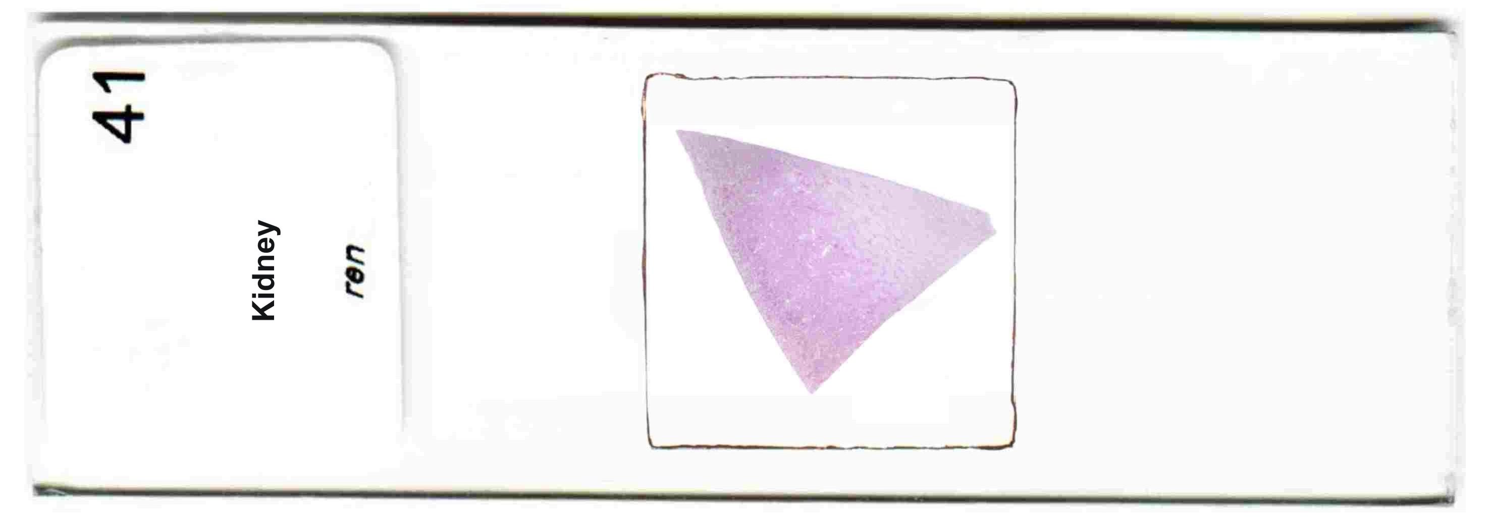
• Kidney
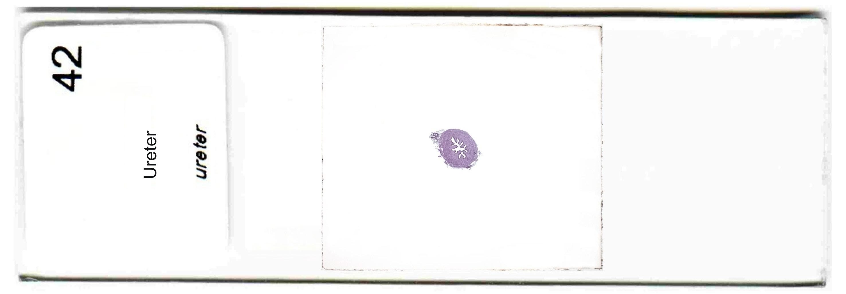
• Ureter
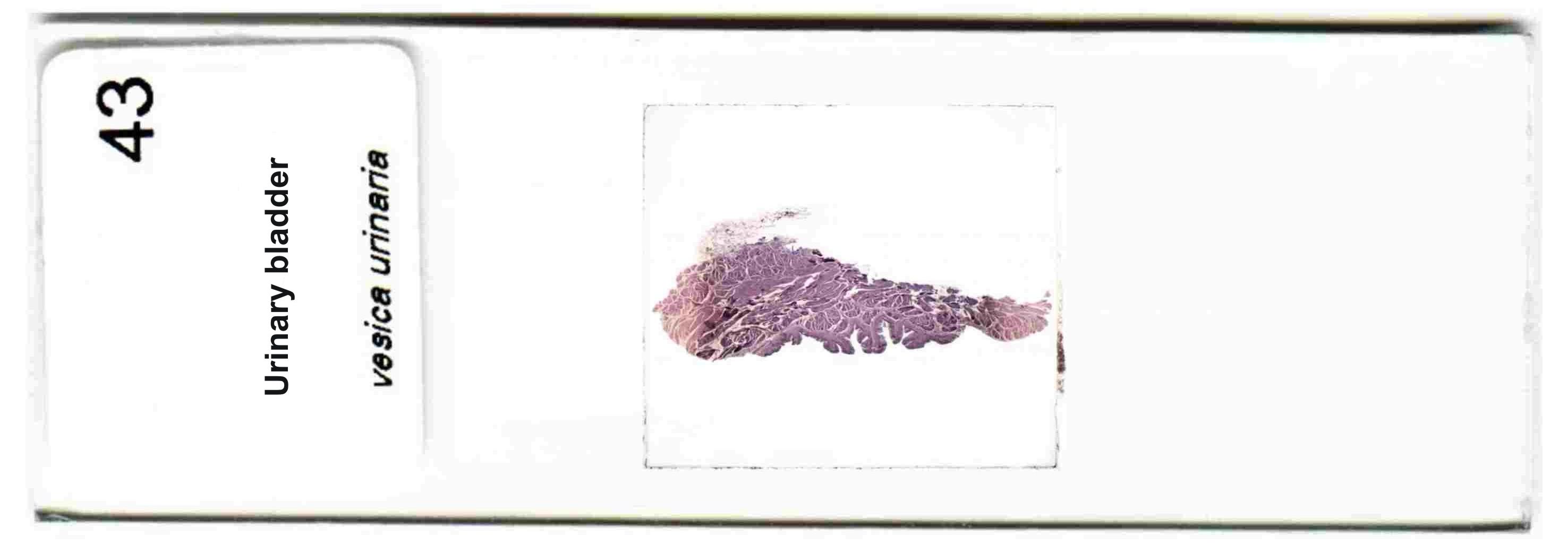
• Urinary bladder
Knowledge of this topic and of the slides is required for revision tests, credit and final examinations in histology and embryology.
-
This part is devoted to light microscopy of the early placenta.

-
This part is devoted to light microscopy of the full-term placenta.

-
This part is devoted to light microscopy of the kidney.

-
This part is devoted to light microscopy of the ureter.

-
This part is devoted to light microscopy of the urinary bladder.

-
Electronic course is supplemented by material for printing and test.
References:
Čihák, R. Anatomie 3, Grada Publishing, Prague, 2004, 692 pp.
Klika, E. Histologie. Avicenum, Prague, 1985, 612 pp.
Krstic, R. V. Human microscopic anatomy. Springer-Verlag, Berlín, 1991, 616 pp.
Mokrý, J., Mazurová, Y., Mráz, J., Čížková, D., Hrebíková, H., Soukup, T. Handbook of Practical Classes in Histology and Embryology, Powerprint, Prague, 2017, 136 pp. -
