Kidney
Theory and slide.
1. DESCRIPTION
Kidney (its exocrine portion) has a structure of compound tubular gland; main function is to produce the urine. Principal structure is composed of renal tubules. Nephrons (approx.. two millions) form the initial part of the tubules (approx. 2 cm in lenght), very short connecting piece. These parts have the same origin; they are derived from metanephrogenic blastema. Other parts of renal tubules, which participate in concentration of the urine include collecting and papillary ducts.
Kidney parenchyma is divided in cortex and medulla.
RENAL CORTEX
A detailed description is provided in the lecture. Principal components of the cortex include: Renal corpuscle (corpusculum renis Malpighi) is the most conspicuous structure of the kidney (Fig. 1 B) composed of the glomerulus (tuft of fenestrated capillaries) invested by a double-layered Bowman's capsule. The layers of Bowman's capsule are separated by the urinary Bowman's space, which collects a primary urine that is drained in the proximal tubule.
Proximal tubule (Fig. 1A) occurs in the cortex in the vicinity to renal corpuscles. It is lined by simple columnar epithelium called nephrocytes. Nephrocytes are relatively large. A luminal surface contains a brush border formed by high microvilli of irregular lengths; as a result the lumen is irregular and narrow (Fig. 2 C).
Distal tubule (Figs. 1A and 2) is lined by smaller nephrocytes with rare short microvilli. For that reason, the lumen is a broad and of a regular shape. A site that comes in contact with a vascular pole of the corresponding renal corpuscle forms the macula densa, which is a component of the juxtaglomerular apparatus responsible for a major endocrine function of the kidney (see ELC „Endocrine system“).
 |
 |
|
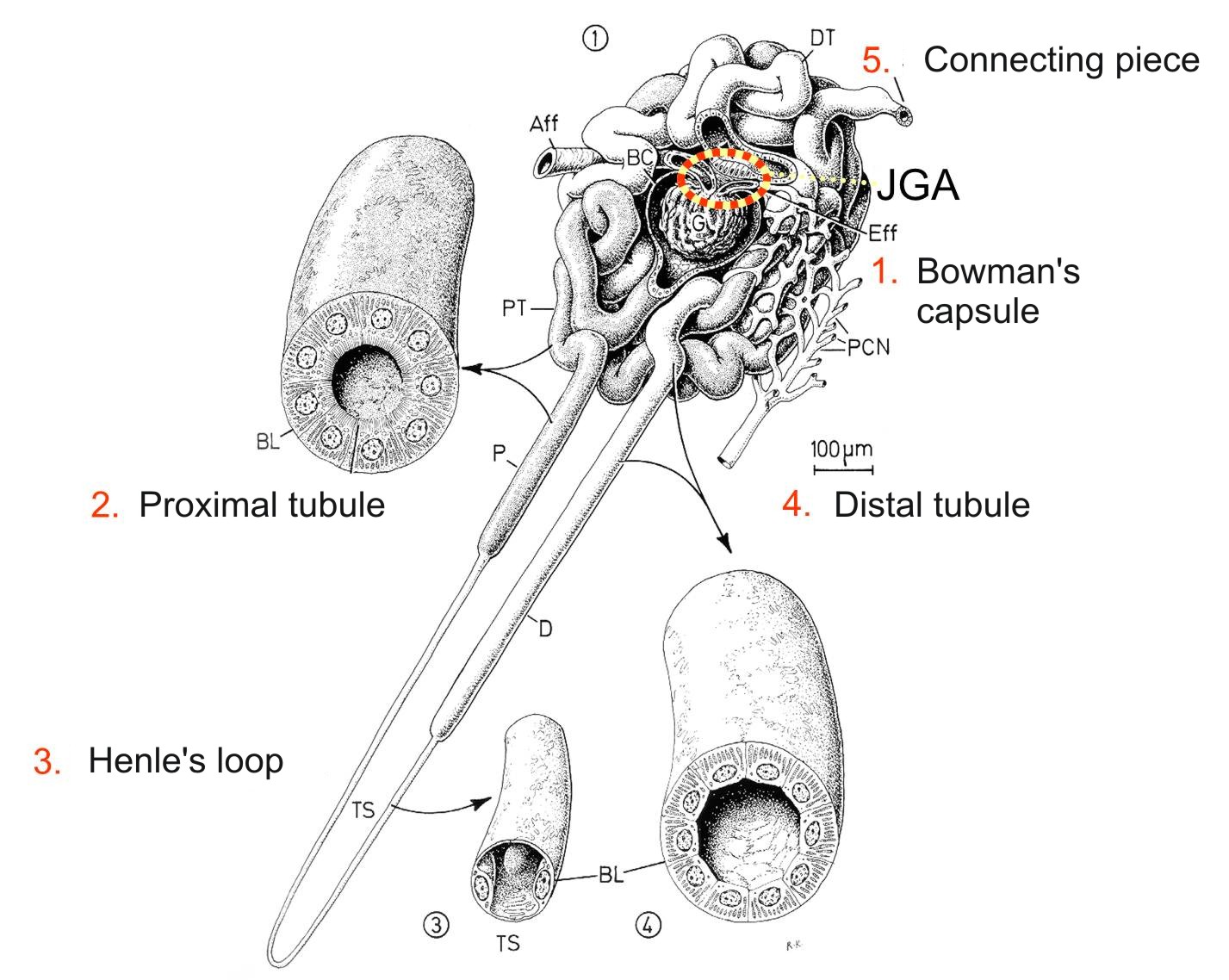
| Fig. 1A. 3D reconstruction of nephron. Parts of nephron are labelled with red numbers. Aff, afferent arteriole; Eff, efferent arteriole; BC, Bowman's capsule JGA, juxtaglomerular apparatus; PT, proximal tubule, stočená část; DT, distal tubule, convoluted part; P, proximal tubule, right part; D, distal tubule, right part; TS, thin segment of Henle's loop; BL, basal lamina; PCN, peritubular capillary network. Krstic, R. V. Human microscopic anatomy |
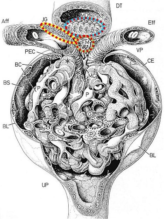
Fig. 1B. 3D reconstruction of renal corpuscle. Aff, afferent arteriole; Eff, eferent arteriole; BC, Bowman's capsule; JG, juxtaglomerular cells; VP, vascular pole; DT, distal tubule, convoluted part; P, proximal tubule, right part; D, distal tubule; UP, urinary pole BL, basal lamina; G, glomerulus; P, podocyte; MC, mesangial cells; EM, extraglomerular mesangial cells; CE, external layer of Bowman's capsule. Krstic, R. V. Human microscopic anatomy |
|
|
RENAL MEDULLA Henle's loop (Fig. 1A) enters the medulla as a descending loop and then turns up as an ascending loop. In a microscope we can distinguish a thin segment (15 μm) lined by a simple squamous epithelium and a thick segment (30 μm) lined by simple cuboidal epithelium that is continuous with a distal tubule. |
||
 |
||
| Fig. 2. 3D reconstruction of distal tubule. A corresponding part of the nephron (A) is indicated in a square. A lining epithelium is formed by nephrocytes (B) with spherical nuclei (N) and basal labyrinth (lab). Basal lamina is labelled in pink colour. A scheme of ligh microscopic structure of proximal (C) and distal (D) tubule depicts crucial structural differences. Krstic, R. V. Human microscopic anatomy |
||
|
The other tubules that are derived from the ureteric bud include collecting and papillary ducts. Collecting duct is lined by a simple cuboidal to columnar epithelium; the cytoplasm is very pale and boundaries between the cells are well visible and apical cytoplasm bulges into the lumen. Papillary ducts (of Bellini) lined by simple columnar epithelium collect the urine from 5-7 collecting tubules; however, in our sides these structures are not included. |
||