Ureter
Theory and slide.
1. DESCRIPTION
Ureter is a tubular organ of urinary passages with length over 25 cm that transports urine form the renal pelvis to the urinary bladder. A wall of the ureter consists of three layers: i) Tunica mucosa, ii) Tunica muscularis and iii) Tunica adventitia.
The mucosa is lined by the urothelium (classified as a pseudostratified cuboidal epithelium). This epithelium is also referred as to transitional epithelium. Lamina propria mucosae is formed by a loose connective tissue with many elastic fibres, which permits a formation of longitudinal mucosal folds well visible in a transverse section.
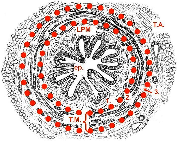
Fig. 1. Scheme of ureter in a tranverse section. Mucosa consists of lamina epithelialis (ep.) and lamina propria mucosae (LPM). T. muscularis is formed by three layers of smooth muscle (1., 2. and 3.) with different orientation. T. adventicia (T.A.) is located externally.
Klika, E. Histologie
T. muscularis is formed by a smooth muscle. In upper two thirds t. muscularis consists of inner longitudinal and outer circular layers. In the lower third of the ureter, an additional longitudinal outer layer is apposed. Layers of smooth muscle are responsible for peristaltic movement.
An outer layer is formed by t. adventitia of loose connective tissue (ureter is located retroperitoneally).