Urinary bladder
Theory and slide.
1. DESCRIPTION
A wall of the urinary bladder consists of i) Tunica mucosa, ii) Tunica muscularis and iii) Tunica adventitia / serosa.
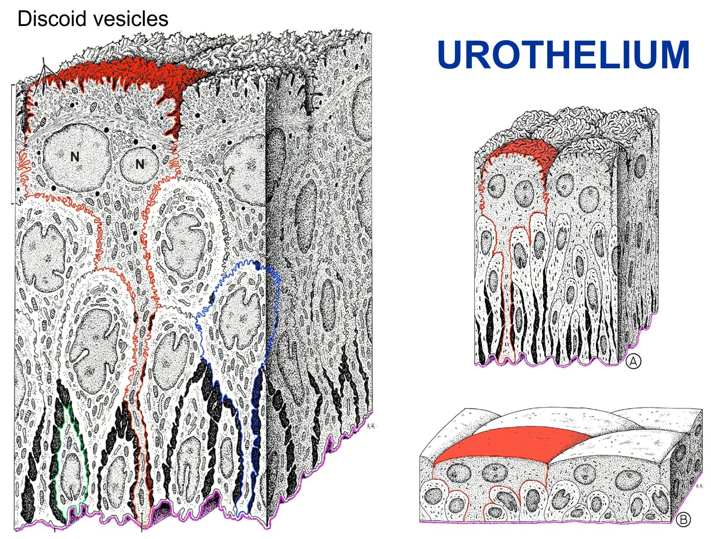
The mucosa is lined with the urothelium (classified as a pseudostratified cuboidal epithelium). This epithelium is also called as the transitory epithelium because its structure changes according to its content: an empty bladder has 6-8 cell rows while a full one has only 2-3 cell rows. The lamina propria mucosae under the basal lamina is formed by a loose connective tissue and permits to form mucosal folds. A smooth mucosa in the trigonum vesicae facilitates removal of urine into the urethra.
 |
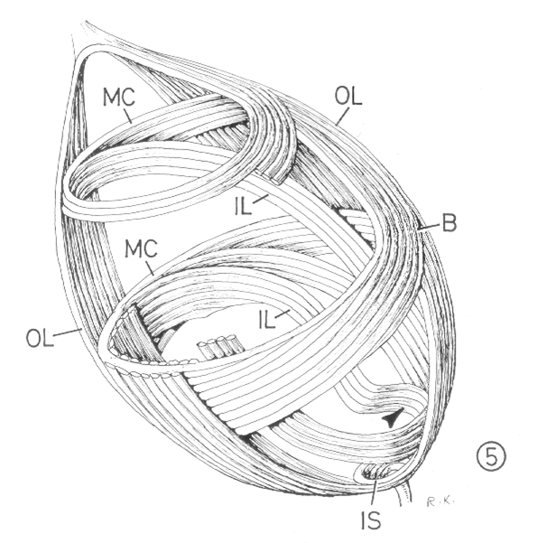 |
Fig. 1. 3D reconstruction of mucosa and muscle of urinary bladder. The urothelium (left) is lined with several cell layers: basal cells (green) are attached to the basal lamina (magenta), middle cells (blue) have a shape of tennis racket while superficial cells (red) have a cuboidal shape but they are still anchored to the basal lamina with their cytoplasmic process. The apical plasmalemma is very thick, the cytoplasm contains discoid vesicles that fuse with the plasmalemma when the bladder is extended. Figs. A and B show changes occurring after extension of the urothelium. An image on right show a complicated arrangement of muscle layers inside of the wall of the urinary bladder.
Krstic, R. V. Human microscopic anatomy
The t. muscularis consists of a smooth muscle, which is arranged in three layers: an inner layer is plexiform, middle circular (the thickest part constitutes the musculus sphincter vesicae) and outer is longitudinal. A musculature of all three layers functions as the musculus detrusor, i.e. it is important in emptying the bladder.
From outside the bladder is covered mostly by t. adventitia of a loose connective tissue; a posterior aspect is covered by the serosa.