DORSAL ROOT GGL.
1. DESCRIPTION
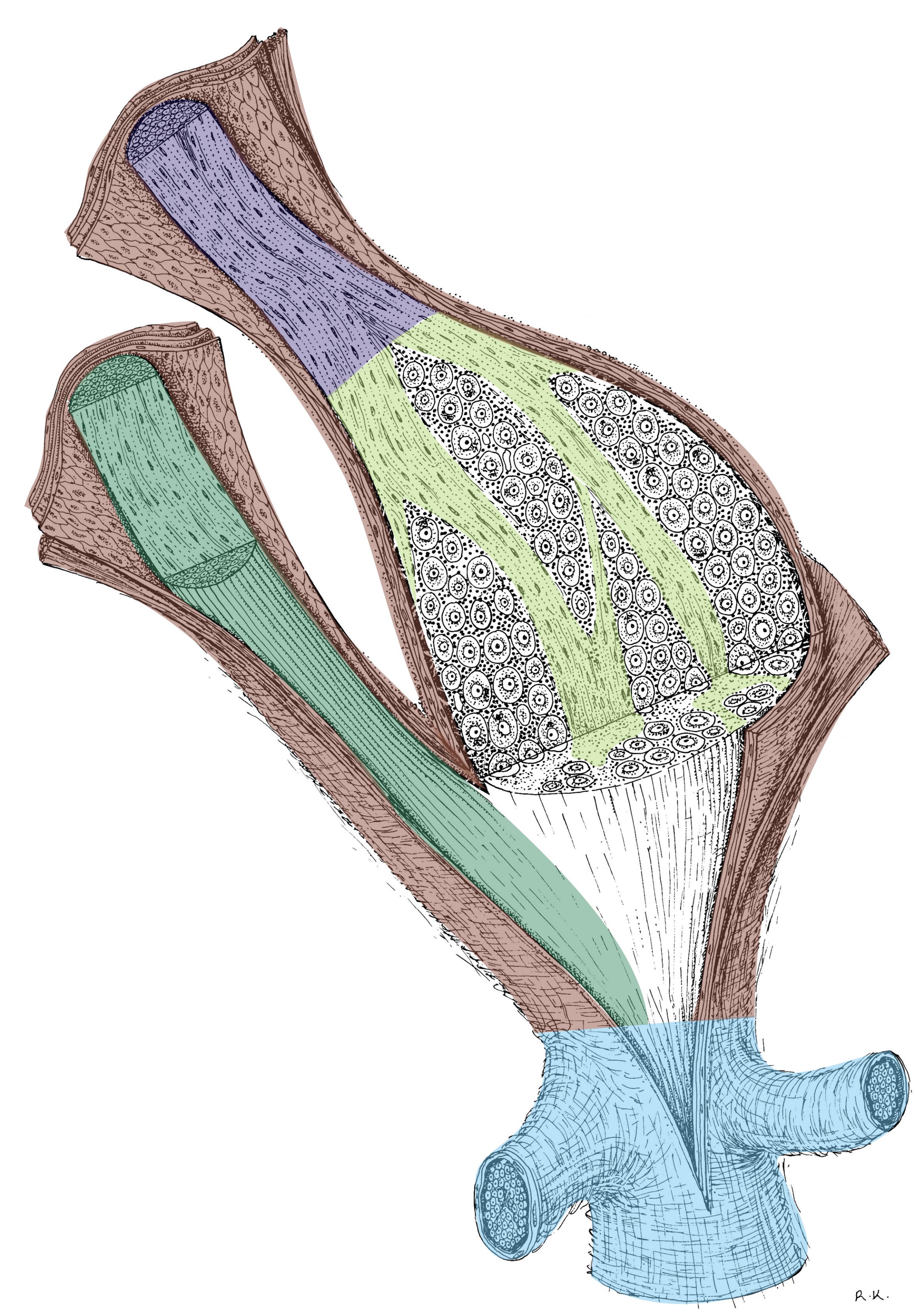
 Dorsal root ganglion arises from accumulation of pseudounipolar neurons in the area of the foramen intervertebrale. Dorsal root ganglion (Fig. 1) is inserted in the dorsal spinal roots (purple). A surface is covered by a capsule (brown), which is a continuation of the coverings of the spinal cord; the most conspicuous is the outermost layer of the dense connective tissue. Fusion of ventral (dark green) and dorsal roots (purple) gives rise to the spinal nerve (indicated in light blue). Under the capsule, the bodies of ganglionic cells are clustered in groups (white). A typical feature of the dorsal root ganglion is a regular arrangement of its components, i.e., the perikarya are not mixed with nerves. Nerve fibres (light green) are aggregated and fill in the spaces between groups of nerve bodies.
Pseudounipolar neurons of dorsal root ganglion have large perikarya reaching a size 100 μm. A common process leaving the perikaryon branches in a shape of a letter "T". The perikaryon is ensheathed by a single layer of satellite cells (amphicytes). Due to a single process and a large perikaryon there are many satellite cells forming a continuous layer. Pseudounipolar neurons send their afferent arm that accepts sensitive/sensory information from the periphery (e.g. nociception, proprioreceptors etc.). Their efferent arm brings information in the spinal cord via dorsal roots to synapse with spinal neurons - for that reason there are no synapses inside of the dorsal root ganglion. Nerve fibres of these ganglionic cells are myelinated.
Cerebral ganglia show a very similar structure. These ganglia also contain pseudounipolar ganglion cells with the exception of the ggl. vestibulocochleare, which is the only ganglion containing bipolar neurons.
Fig. 1. 3D reconstruction of dorsal root ganglion. Illustration legend can be found in the text.
Author: R.V: Krstic; colours: J. Mokrý
