SPINAL CORD
1. DESCRIPTION
The dorsal spinal cord (medulla spinalis) is the oldest part of the CNS. It consists of a column of nervous tissue about 40-45 cm long, which is located in the vertebral canal. A cross-section of the spinal cord has a characteristic appearance - the grey matter is centrally located and butterfly-shaped (Fig. 1A), with the central canal running through its centre, lined by the ependymal cells and containing cerebrospinal fluid. The anterior horns are relatively broad, do not reach the edge of the spinal cord, and contain perikarya of the largest nervous cells, the so-called alpha motor neurons. The posterior horns of the spinal cord are slender and often reach the edge of the spinal cord. The grey matter is surrounded by white matter, which is composed mainly of myelinated nerve fibres arranged in anterior, posterior and lateral fascicles. The anterior median fissure, which is filled by the sparse loose connective tissue of the pia mater, is pressed into the ventral surface of the spinal cord. At the end of this fissure along the spinal cord runs the anterior spinal artery, which provides nutrition to the spinal cord. The posterior fascicles overlie the thin dorsal median septum.
Basic orientation in slide of the spinal cord, including identification of motor neurons, is explained in the Practical classes "Nervous System".
 |
 |
|
|
Fig. 1 A. Transverse section of the spinal cord. A scheme shows structures visible in the transverse section of the spinal cord. Ventral horns contain perikarya of alpha motor neurons. The subarachnoid space contains ventral and dorsal roots and accompanying blood vessels. |
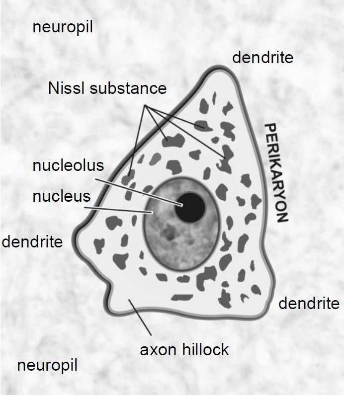
Fig. 1 B. Terminology used in neuron description. A scheme shows structures visible at light microscopy in a paraffin-embedded section of the ventral horns of the spinal cord containing alpha motor neurons.
Mokrý, J. et al. Handbook of Practical Classes in Histology and Embryology. |
The spinal cord, like other compartments of the CNS, is covered by the meninges. The pia mater covers the surface of the spinal cord and its connective tissue passes as an endoneurium between the nerve fibres of the ventral and dorsal roots. The arachnoid membrane covers a surface of the nerve bundles as the perineurium when the spinal roots are spaced apart. The dura mater, consisting of dense connective tissue, passes over the spinal nerves as the epineurium.
 |
 |
|
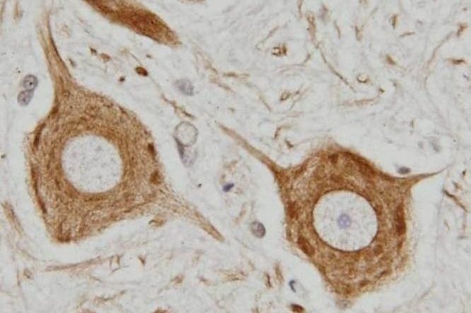
| Fig. 2 A. Imunohistochemical staining of neurofilaments. Perikarya of motor neurons and their cytoplasmic processes in the neuropil can be specifically visualized by peroxidase immunohistochemistry (brown). The microphotograph demonstrates that motor neurons are multipolar. The nuclei were counterstained with hematoxylin. Author of microphotograph: Jaroslav Mokrý. |
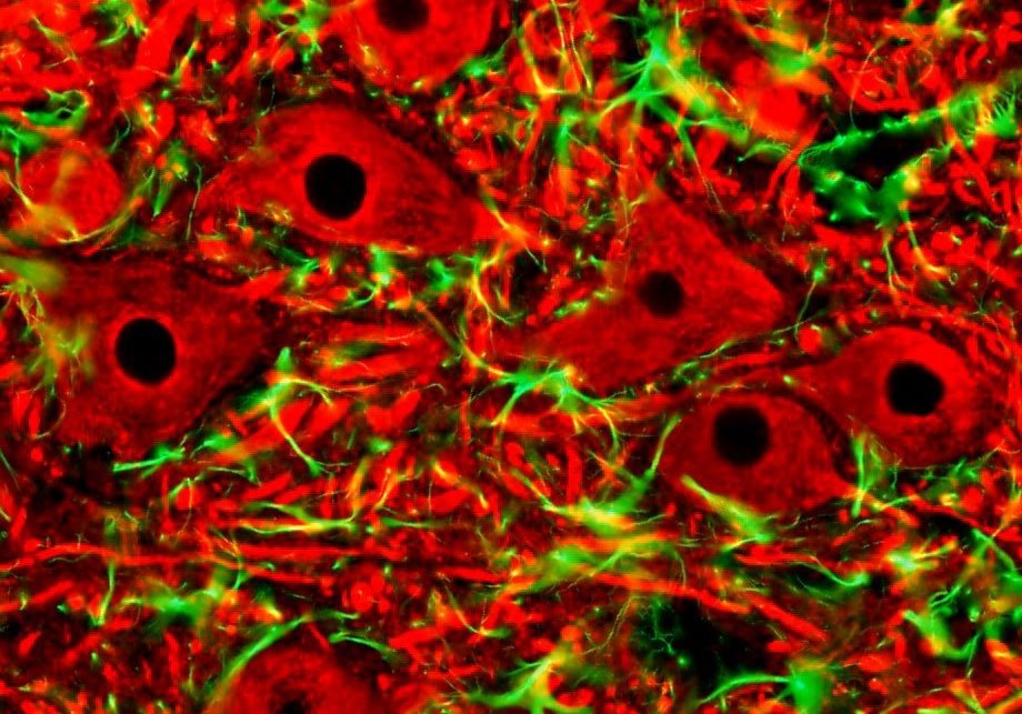
Fig. 2 B. Double immunofluorescence. Using specific antibody against pan-neurofilaments (red) and GFAP (green), motor neurons and astroglia of the spinal cord ventral horn are labelled in distinct colours. Cell nuclei do not contain intermediate filaments and therefore remain dark. Author of microphotograph: Jaroslav Mokrý. |