Ureter
Theory and slide.
2. SLIDE: URETER
|
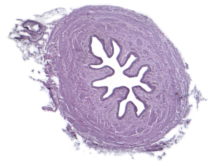
ORIENTATION IN THE SLIDE A general structure of the ureter was described on previous page. In a transverse section it appears as a tine tubular organ (thickness of the wall is approx. 1 mm). A wall consists of three layers: i) T. mucosa is lined with the urothelium; a loose connective tissue in the lamina propria allows formation of longitudinal foldings (three mucosal folds are shown in green). Presence of foldings gives rise to a stellate-like lumen (indicated by asterisks). ii) A middle layer is t. muscularis of smooth muscle. The upper portion consists of two layers; in a low third the third layer is added). An inner layer is oriented longitudinally (indicated in red); another layer is circular (purple). The third layer is formed by longitudinally running cells. iii) The outermost layer, t. adventitia (yellow), contains a loose connective tissue with blood vessels, nerves and adipocytes. |
Fig. 2. Interactive slide of ureter: Low magnification. Moving a cursor to an image shows structures in the slide in different colours. Illustration legend can be found in the text in left. |
|
Ureter wall Longitudinal mucosal folds of the ureter are lined by the urothelium. A core of the folds is formed by a loose connective tissue constituting the lamina propria mucosae (in green). A mucosal connective tissue is locally infiltrated by lymphocytes (purple) that can form rare lymphatic nodules. Under the mucosa there is the tunica muscularis consisting of the smooth muscle. An inner layer is oriented longitudinally; in a transverse section, the bundles of this layer are oval (red) – only he largest spherical diameters of myocytes reveal the nucleus. In a next layer, the smooth muscle is arranged circularly; in a transverse section, spindle-shapes myocytes can be seen containing elongated rod-like nucleus in the central part. Fig. 3. Interactive slide of the ureter wall: A detailed view. Moving a cursor to an image shows structures in the slide in different colours. Illustration legend can be found in the text in left. Haematoxylin-eosin staining. |
 |
|
The covering epithelium lining the ureteral mucosa (urothelium or pseudostratified cuboidal epithelium) has a characteristic appearance. The epithelium is relatively thick and consists of several rows of cells. The basal cells are most often triangular (shown in green) and are attached to the basal lamina (its course is shown in purple). Other cells are longer, and their nuclei are located higher up. Most typical are the tennis racket-shaped cells (blue), which do not reach the surface with their apical pole. The most differentiated cells line the surface (red); these cells are roughly cuboidal in shape and therefore have a round nucleus. Some surface cell nuclei are shown in black. The apical membrane of the surface cells of the urothelium is thickened to resist the action of urine. The apical cytoplasm contains discoid vesicles; these structures are visible in light microscopy as the crusta, which is visible as a dark line, the course of which is shown as a grey line. Fig. 4. Epithelium of the ureter: A detailed view. Moving a cursor to an image shows structures in the slide in different colours. Illustration legend can be found in the text. |
 |