EYELID
This part of the E-course is devoted to a microscopic structure of the eyelid.
1. DESCRIPTION
The eye is protected by the eyelids, which has the character of a cutaneous fold. The eyelid is reinforced by a tarsal plate made of a dense connective tissue, which contains about 25 Meibomian sebaceous glands. The tarsal plates are attached to the Müllerian smooth muscle of the superior and inferior tarsal muscles. The eyelids also contain the skeletal muscle (palpebral part of the orbicular oculi muscle); the sphincter of the eyelid closes the eye slit periodically during blinking. The levator palpebrae superioris that elevates the upper eyelid (it is inserted at tarsus and subcutaneous connective tissue) it is not seen in the slide. The outer surface of the eyelids is covered by the fine skin, the inner concave surface is covered by the conjunctiva. The conjunctiva is lined by a stratified columnar epithelium in which goblet cells are scattered. The eyelid conjunctiva extends far and turns into the bulbar conjunctiva at the fornix where the mucous cells are arranged in small endoepithelial glands; the conjunctiva here up to the limbus is freely movable so as to allow the movements of the eyeball. At the junction of both surfaces of the eyelid is the intermarginal zone, in which cilia are inserted in 3-4 rows. Zeiss sebaceous glands and Moll's apocrine sweat glands are also associated with the eyelashes. Behind the eyelashes, the Meibomian glands arranged in one row open; their secretion, rich in fat, prevents the tears to overflow onto the face.
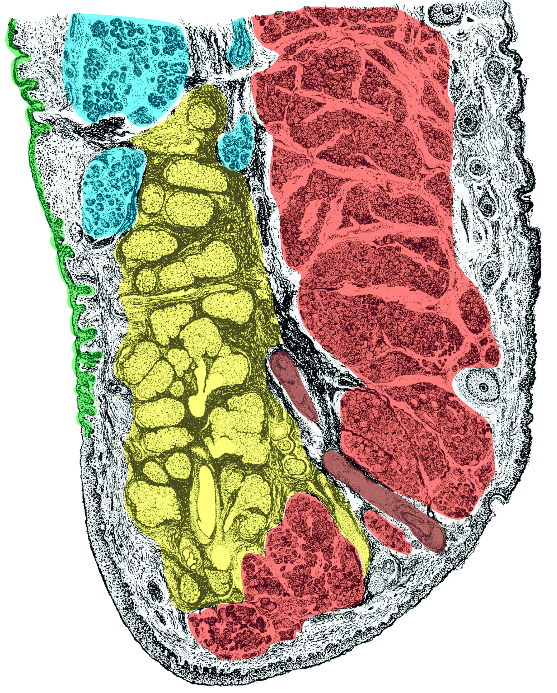 |
Fig. 1. Schematic drawing of the upper eyelid. The tarsal plate is marked in yellow; the Meibomian gland can be seen inside. The orbicularis oculi muscle is coloured red, cilia brown, and lacrimal gland blue. The inner surface is covered by the conjunctival epithelium (green), while the outer surface (lower and right edge) is covered by a thin type of the skin. Image taken from E. Klika et al. Histologie, colouring: Jaroslav Mokrý |
|
The lacrimal gland is also related to the eyelid, as it is located in the outer upper quadrant of the orbit and is divided into an eyelid and an orbital part by the tendon of the upper eyelid elevator. The ducts of the gland exit laterally into the superior fornix, and the lacrimal fluid flows down the eyeball into the lacrimal lake (lacus lacrimalis), from which it drains through the two puncta lacrimalia into the lacrimal canaliculi and then into the lacrimal sac (saccus lacrimalis), whence it drains into the lower part of the nasal cavity through the nasolacrimal (tear) duct. Tears not only moisten the cornea and allow the eyelids to glide easily, but also have an antimicrobial function.