POSTERIOR SEGMENT OF EYE AND OPTIC NERVE
This part of the E-course is devoted to a microscopic structure of the posterior segment of the eye and the optic nerve.
1. DESCRIPTION
The structures of the posterior segment of the eye are shown in Figure 1A and include the optic portion of the retina, choroid, and sclera (each is a derivative of a different layer). Also visible is the optic nerve (in longitudinal section), protruding from the posterior portion of the eyeball. The sclera was described in the previous slide (anterior segment of the eye). The choroid as a derivative of the vascular coat contains loose connective tissue with a large number of blood vessels and pigment cells. Four layers of the choroid can be distinguished in the section, which are described below.
 |
 |
|
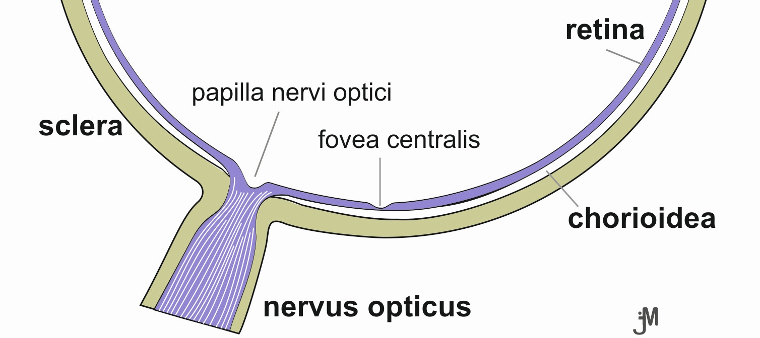
| Fig. 1A. Structures of the posterior segment of the eye. In the meridional section of the right eye, the sclera is marked in green, the retina in violet, and the choroid between them in white. Author of image: Jaroslav Mokrý |
Fig. 1B. Arrangement of neurons in the retina. Rods (gray) and cones (red) are connected to bipolar neurons (black), which relay the signal to ganglion cells (orange). Müller cells are stained in blue and the pigment epithelium in brown. |
|
|
The most important structure is the retina, or its optic part. It is a derivative of the nervous coat, and therefore in its laminar arrangement we can find nervous cells - specifically the first 3 neurons of the optical pathway. The pars optica retinae extends from the ora serrata to the papilla of the optic nerve and shows a characteristic laminar arrangement. The outer, i.e., choroid-facing surface is covered by the pigmented epithelium, which is in contact with the outer segments of the rods and cones (Fig. 1B). The photoreceptor bodies, as well as the other retinal layers with which these cells are in contact, are arranged toward the interior of the eyeball filled with the vitreous body. Thus, a photon carrying image information must pass through all the retinal layers on its path before it reaches the photosensitive discs of rods and cones. The first neuron is modified into photosensitive cells, rods and cones. The excitation produced by the photon impact is transmitted by the axon to the bipolar neurons, which pass the already extensively processed signal to the multipolar neurons (ganglion cells). The vertical interconnection of three neurons serves to partially process the image information already at the retinal level and at the same time reduces the number of axons conveying information from the eye to the CNS by about 100 times (108 photoreceptors, but only 106 fibres in the n. opticus). Neurons within the retina are also interconnected horizontally via horizontal and amacrine cells (neurons). The part of the retina that lies in the visual axis of the eye is predisposed for the sharpest vision. This area of the retina is slightly yellow-green in colour and is therefore referred to as the macula lutea; a small pit (fovea centralis maculae), about 1/3 mm in size, is recognisable by the ophthalmoscope. The fovea contains mainly cones, which mediate colour vision. The bipolar and ganglion cells, with which the cones of the macula are associated, are displaced to the side (creating a small depression in the retina), so that photons can fall directly on the photoreceptors in the area of sharp vision. Unmyelinated ganglion cell axons are collected from all parts of the optic retina and directed to the papilla of the optic nerve. The image that reaches the papilla cannot be perceived because this area of the retina lacks photoreceptors (blind spot). Here, all the nerve fibres of the ganglion cells converge, together penetrate the wall of the eyeball and form the optic nerve (nervus opticus); the fibres acquire a myelin sheath only after they have penetrated the sclera. |
||