ANTERIOR SEGMENT OF THE EYE
This part of the E-course is devoted to a microscopic structure of the anterior segment of the eye.
1. DESCRIPTION
The eye is the most important sensory organ that conveys our vision. Through the eye, we perceive the image of the surrounding world through light that is projected onto the light-sensitive retina. In terms of its structure, the eye is the most complex sensory organ. The wall of the eyeball is made up of three concentric layers. The outer layer, the fibrous coat, differentiates into the sclera and cornea. The middle layer of the eyeball wall is the vascular coat (uvea). Uvea comprises three components - the choroid, the ciliary body, and the iris. Uvea is a layer composed of loose connective tissue containing numerous blood vessels and melanocytes. The pigment cells, together with the retinal pigment epithelium, form a black chamber inside the eye that prevents light reflection. In the inner layer of the eyeball, the nervous coat, lies the retina. Its anterior part covering the ciliary body and the posterior surface of the iris is double-layered and forms the blind part of the retina, as it lacks photoreceptors and nervous cells. The optic part of the retina extends from the ora serrata to the papilla of the optic nerve. The optic nerve fibres convey information from the eye to the brain for further processing.
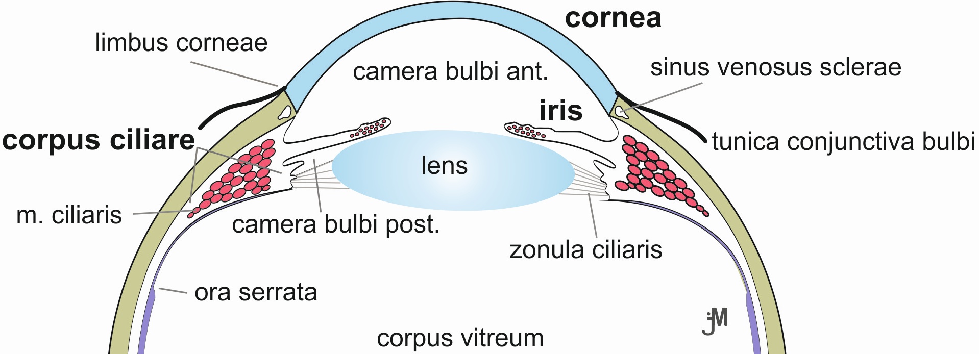
Due to the size of the eye, we present the tissues of the eye on two slides; the lens and vitreous body are usually damaged and removed during processing. The structures of the anterior segment of the eyeball are indicated in Figure 1; the most important structures include the cornea, the sclera, the ciliary body, the iris, and the blind portion of the retina.
The cornea is the transparent anterior part of the eye, which is bulging forward. Five distinct layers can be distinguished in its histological section. The outer surface is covered by the anterior corneal epithelium (Fig. 1), which consists of a stratified squamous non-keratinized epithelium located on a well-defined Bowman's membrane (an underlying acellular structure composed of the basal lamina and adjacent collagen fibrils embedded in the ground substance). The most prominent layer is the corneal stroma composed of many layers of lamellae of collagen microfibrils arranged in parallel (= regular dense connective tissue). Between the lamellae are numerous modified fibrocytes called keratocytes. For the sake of its transparency, this layer is avascular. The posterior surface of the cornea is covered by a simple squamous epithelium, called the posterior epithelium of the cornea whose cells are connected by desmosomes and tight junctions and transport fluid from the corneal stroma. The posterior epithelium sits on Descemet's membrane. The cornea transitions to the sclera at its margin, referred to as the limbus. The histological structure of the cornea changes here (the area of Vogt's palisades is described in detail in the lecture). The sclera occupies the posterior 5/6 of the surface of the eyeball. It is a structurally and functionally supportive tissue consisting of dense connective tissue consisting of parallelly arranged, but crossing, flat bundles of collagen fibres and only a limited number of fibroblasts and ground substance. Because of the minimal number of blood vessels, this part of the eye appears white. The anterior part of the sclera is covered by the conjunctiva lined by the stratified columnar epithelium.
 |
 |
|
| Fig. 1A. Structures of the anterior segment of the eye. Transparent structures are shown in blue, muscles in red, sclera in green and retina in purple. Author of image: Jaroslav Mokrý |
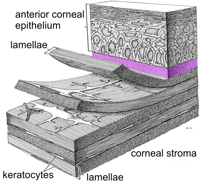
Fig. 1B. Three-dimensional reconstruction of the anterior part of the cornea. The anterior epithelium sits on a thick Bowman's membrane, which is outlined in purple. The collagen microfibrils within the lamellae in the stroma of the cornea are arranged in a parallel pattern; adjacent lamellae cross against each other at nearly right angles. Krstic, R. V. Human microscopic anatomy (colouring: J. Mokrý) |
|
|
The anterior part of the uvea is transformed into the ciliary body (corpus ciliare) and the iris. In a longitudinal section of the eyeball, the uvea expands in the area of the ciliary body and acquires a triangular shape, which is caused by the presence of the smooth muscle (ciliary muscle). The contraction of the ciliary muscle relaxes the tension of the suspensory fibres of the lens, which thus becomes curved and focuses on near subjects. The ciliary muscle is innervated by parasympathetic fibres of the oculomotor nerve of the Edinger-Westphal nucleus after coupling to the postganglionic neuron in the ganglion ciliare. The surface of the ciliary body is covered by the blind portion of the retina (pars ciliaris retinae), formed by two layers of cells. The outer layer adjacent to the ciliary body corresponds to the pigment epithelium, while the inner layer does not contain pigment. The posterior surface of the ciliary body is flat (pars plana), while the ciliary processes, processus ciliares, after which this body was named, are located in the anterior pars plica. The projections are richly vascularized; by ultrafiltration, ion and fluid transport, the secretory activity of the non-pigmented epithelium produces a humor aqueus that serves to nourish the avascular structures of the eye, to remove catabolites and to maintain the shape of the eyeball. The anterior part of the uvea, extending from the corpus ciliare, detaches from the sclera to form the iris. The iris has the appearance of a thin circular disc with a circular opening in the middle (pupil). The size of the pupil is regulated by two muscles: the pupillary sphincter (m. sphincter pupillae) and the pupillary dilator (m. dilatator pupillae), which regulate the amount of light reaching the retina. The sphincter is located near the pupil and is controlled by the parasympathetic; nerve fibres; the dilator muscle, composed of myoepithelial cells, is located in the posterior border layer and is innervated by the sympathetic nerve fibres. The surface of the iris facing the anterior chamber of the eye is lined by a simple squamous epithelium. The stroma of the iris has the character of a gelatinous connective tissue containing pigmented chromatophores, which, together with the two pigmented layers of the pars iridica retinae, prevent light from entering the eye beyond the pupil. The amount of pigment cells determines the final colour of the iris; if melanin is only contained in the pigment epithelium of the posterior edge of the iris, the eye is blue. The inner layer of the eyeball, the tunica nervosa (nervous coat), contains the retina. In the anterior segment of the eye, an inner surface of the ciliary body and iris consists of two layers and is referred to as the blind part of the retina because it lacks photoreceptors and neurons. |
||