SPLEEN
1. DESCRIPTION
The spleen is the largest lymphatic organ and is primarily used to filter blood. It is a secondary lymphatic organ. It is located in the left hypogastrium, i.e. in the abdominal cavity below the diaphragm. Therefore, its surface is covered by the peritoneum. The stroma of the spleen comprises the capsule, trabeculae and reticular connective tissue (the spleen is a lymphoreticular organ). The capsule contains a dense connective tissue with elastic fibres and some smooth muscle cells - the spleen is expansible and able to change its volume (if necessary, it can expel a large volume of blood to the periphery). Inside the finger-like trabeculae, the trabecular arteries and trabecular veins run. - the vessel wall is greatly reduced to an endothelial lining only, as it is reinforced by the trabeculae wall.
In the splenic parenchyma, two morphologically distinct regions can be distinguished: red pulp and white pulp. The colour of the red pulp is determined by the presence of blood. The red pulp consists of venous sinuses and Billroth cords. Splenic venous sinuses represent an irregularly shaped and richly communicating system of sinusoidal capillaries lined by elongated, spindle shaped endothelial cells oriented parallel to the longitudinal axis of the sinus (their nucleus protrudes prominently into the vascular lumen). The sinusoids are externally supported by a system of perpendicularly arranged processes of reticular connective tissue cells. The reticular fibres run longitudinally being surrounded by a narrow rim of the cytoplasm of the reticular cell protrusions and support the sinuses from outside in the manner of transversely oriented rings. The openings between the endothelial cells lack a basal lamina. Sinuses are drained into the venous bed (they are emptied into the veins of the pulp, which are typical venules). Billroth cords are formed by a 3-D meshwork of reticular connective tissue (reticular connective tissue cells and reticular fibres) that fills the spaces between adjacent sinusoids.
 |
 |
|
|
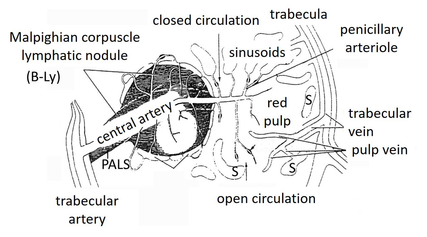
Fig. 1A (left). Scheme of vascularization of the splenic parenchyma. Branching of vessels that participate in open circulation through the parenchyma (i.e. Billroth cords) of the spleen. Lymphatic tissue accumulates in a perivascular space of central artery which forms both components of the white pulp: Malphigian corpuscles and periarterial lyphatic sheaths (PALS). Fig. 1B (right). Schematic drawing of the red pulp. Simplified structure of venous sinusoids (in longitudinal and transverse sections) and Billroth cords (containing a reticular connective tissue). |
||
The white pulp occupies only about 5-20% of the spleen. It consists of lymphatic tissue arranged around the central artery. i) Periarterial lymphatic sheath (PALS) is formed by sleeves of lymphatic tissue oriented along central arteries/arterioles; this tissue contains mainly T-lymphocytes and is therefore referred to as the T-dependent area. ii) Malpighian corpuscles are actually lymphatic nodules and therefore contain mainly B-lymphocytes (B-dependent area of the spleen). There is a marginal zone at the boundary between the white and red pulp, where lymphocytes are transferred from a sinus to the white pulp. The antigen presenting cells (macrophages) are mainly accumulated in this area - this is an area of initiation of the immune response (here blood-borne antigens encounter immunocompetent cells). Detailed structure and functions are described in the lecture “Immune System”.