EYELID
This part of the E-course is devoted to a microscopic structure of the eyelid.
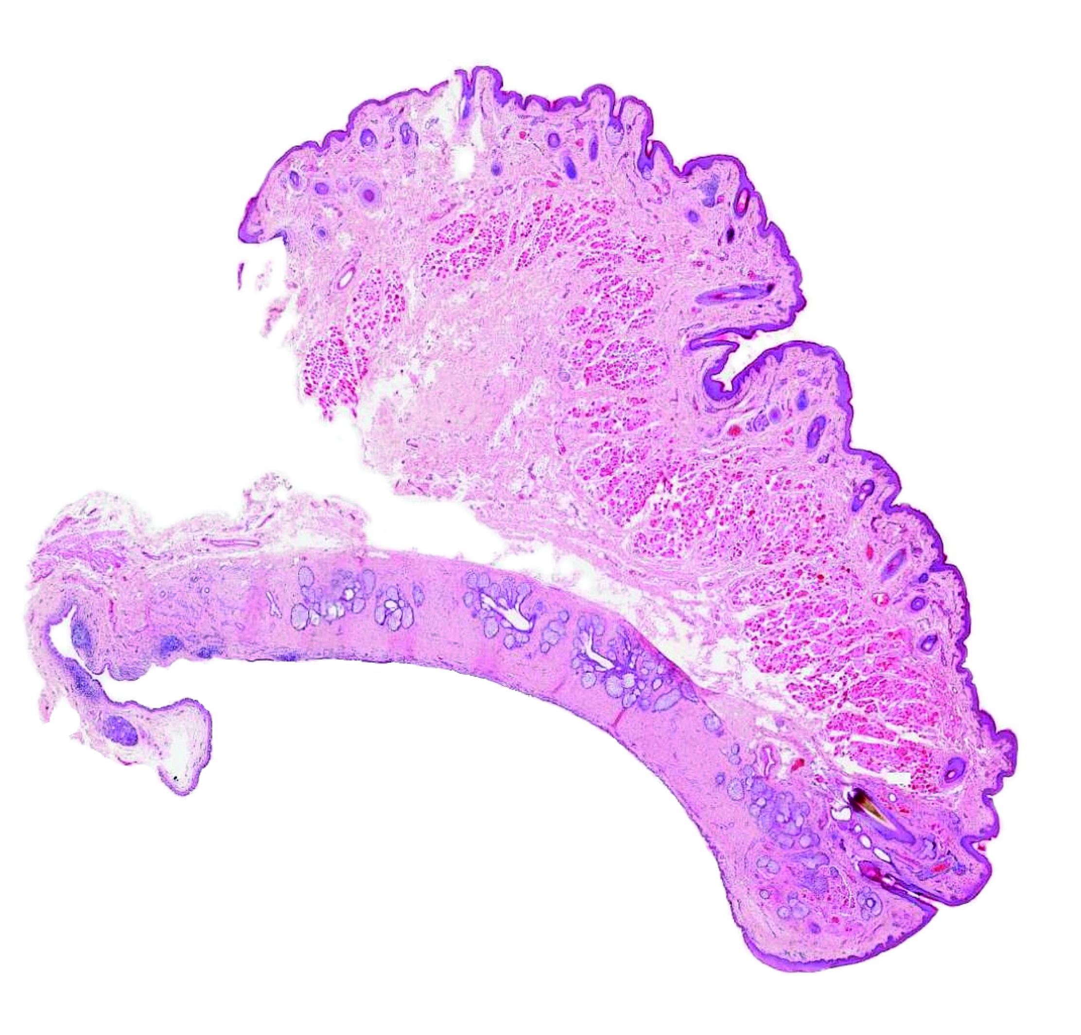
2. SLIDE
|
ORIENTATION IN THE SLIDE On a transverse section of the upper eyelid, two distinct surfaces can be recognised: the skin covers the outer surface, while the inner surface facing the eye is covered by the conjunctiva (marked by the green line). The slide presented here was scanned for the Virtual Atlas and can be viewed in detail in our collection of virtual slides via SmartZoom software. The eyelid contains a rigid connective tarsal plate (yellow) of dense connective tissue near the inner surface, which houses the Meibomian gland. At the upper edge, the smooth muscle (orange) attaches to the tarsus. The conjunctiva near the conjunctival sac contains numerous lymphoid follicles (purple), and occasionally the slide contains an accessory lacrimal gland (blue). In the slide above, the tarsus is artificially detached from the orbicularis oculi muscle (red). A dense connective tissue attached to the tarsus includes the tendon of levator palpebrae muscle (whose muscle tissue is not seen in the slide). Outer surface of the eyelid is covered by a thinly keratinized epidermis, dermis (with dermal adnexa) and hypoderm (subcutis). On the lower margin of the eyelid, the eyelashes (or their large hair follicles) are clearly visible, to which the ciliary glands (Zeiss and Moll's) are attached. Fig. 2. Interactive slide of the eyelid: Survey. Move the cursor over the image to show the basic structures of the slide in a different colour scheme. The legend is given in the text above. Author of microphotography: J. Mokrý. |
|
|
OUTER SURFACE OF THE EYELID The skin surface of the eyelid is lined by a stratified squamous thinly keratinized epidermis (epidermis - shown in orange in interactive Fig. 3). The dermis contains small blood vessels, nerves and cutaneous adnexa, notably small hair follicles (brown) and associated sebaceous glands (not seen in Fig. 3) and eccrine sweat glands (green). A detailed description of the skin glands can be found in the e-course Practical classes 2: Glandular Epithelium. The hypoderm (tela subcutanea) lacks fat cells; skeletal muscle fibres belonging to palpebral part of the orbicular oculi muscle can be found in deeper area. The situation in the figure corresponds to the area indicated by a rectangle 1 in Fig. 2. Fig. 3. Interactive slide of the skin surface of the eyelid: Detail. Move the cursor over the image to show the basic structures of the slide in different colours. The legend is given in the text above. |
 |
|
FREE MARGIN OF THE EYELID The situation depicted in Fig. 4 corresponds to the area indicated by rectangle 2 in Fig. 2. The free margin is covered by thin skin, which contains eyelashes in 3-4 rows. Two of their thick hair follicles are labelled in brown in the longitudinal section. The ciliary glands associated to eyelashes include: sebaceous Zeiss‘ glands (shown in white) and the Moll‘s glands (apocrine sweat glands, in green). A detailed description of the cutaneous glands can be found in the e-course Practical Classes 2: Glandular Epithelium. The smooth ciliary muscle is also seen near the eyelashes in the microphotograph (orange). The outer (ventral) surface of the eyelid (right in Fig. 4) is covered by thin skin, under which is the orbicularis oculi muscle (red - muscle fibres are visible here in a transverse section); the muscle has several parts and therefore its fibres can also be detected near the eyelashes and the tarsal plate. The core of the eyelid contains the tarsal plate (tarsus - yellow) made of dense connective tissue, in which the Meibomian sebaceous gland is found; the alveoli of this gland are marked by a dashed line. Its ducts exit just anterior to the inner margin of the eyelid. The inner lining of the eyelid consists of the conjunctiva. The surface is covered by a stratified columnar epithelium, the course of which is marked by a green line; beneath the epithelium is the lamina propria mucosae of a loose connective tissue. Fig. 4. Interactive preparation of the free eyelid margin: Detail. Move the cursor over the image to show the basic structures of the slide in a different colour scheme. The legend is given in the text above. Author of microphotography: J. Mokrý. |
 |
|
CONJUNCTIVA AND LACRIMAL GLAND The conjunctiva covers the inner (concave) part of the eyelid (palpebral conjunctiva) and passes through the superior and inferior fornix to cover the ventral surface of the eyeball (t. conjunctiva bulbi). The situation in Fig. 5 shows the lining epithelium of the conjunctival sac and corresponds to the part indicated by a rectangle 3 in Fig. 2. The surface of the conjunctiva is covered by a stratified columnar epithelium (green) in which goblet cells are scattered (white) that contribute to the lubricating function of the tear film. The adjacent connective tissue layer is composed of loose connective tissue that contains numerous small blood vessels. Lymphatic follicles, although not shown in the microphotograph, are an integral part of the lamina propria mucosae. The ducts of the lacrimal gland open into the upper fornix; a portion of the lobe of the accessory lacrimal gland (blue) is depicted in the lower part of Fig. 5. The secretory portion consists of alveoli (serous acini), which contain a single layer of serous cells as well as myoepithelial cells. The intralobular ducts lined by simple cuboidal epithelium are embedded in the loose connective tissue of the lobe and drain secretions into the interlobular ducts (lined by a double-layered columnar epithelium). Fig. 5. Interactive slide of conjunctiva and lacrimal gland: Detail. Move the cursor over the image to show the basic structures of the slide in a different colour scheme. The legend is given in the text above. Author of microphotograph: J. Mokrý. |
 |