
SYMPATHETIC GANGLION
2. SYMP. GGL. SLIDE
ORIENTATION IN THE SLIDE
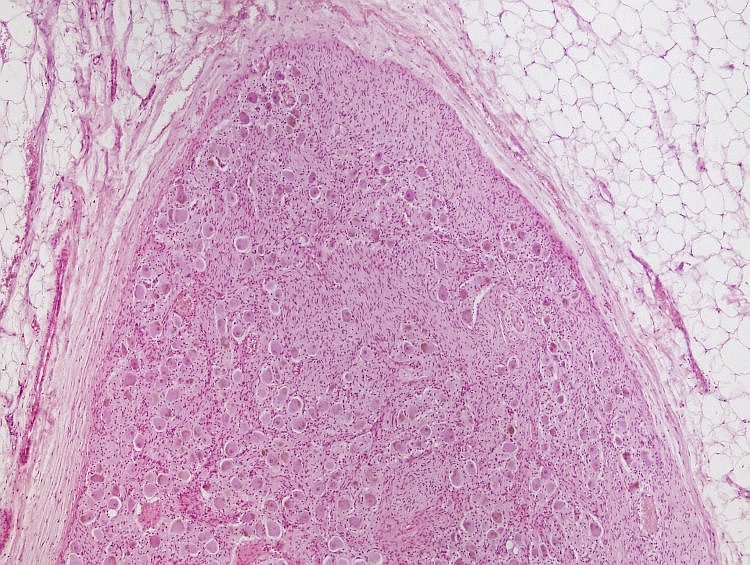
Fig. 2. Interactive slide of the ganglion: A survey. Illustration legend can be found in the text. |
 |
|
|
The parenchyma of the ganglion (Fig. 3) contains well apparent large bodies of ganglionic (nervous) cells (in an interactive slide indicated by a dotted line). Inside of the perikaryon the oval nucleus (grey) with a prominent nucleolus can be seen (black). The cytoplasm contains well visible accumulations of basophilic Nissl tigroid substance (purple) and sometimes also pigment lipofuscin (brown). Many processes of multipolar ganglionic cells are not seen well in a section. A body of the ganglion cell is covered by a discontinuous layer of satellite cells (red) called also as amphicytes. Nerve fibres filling in the space between the ganglionic cells are unmyelinated. Fig. 3. Interactive slide of the ganglion cells. Moving a cursor to an image shows structures in the slide in different colours. llustration legend can be found in the text.Author of the microphotograph: J. Mokrý. |
 |