ENDOTHELIAL PROGENITOR CELLS
Description of endothelial cells, their development and their progenitor cells incl. methods of isolation and usage in stem cell therapy.
7. NESTIN EXPRESSION IN BLOOD VESSELS
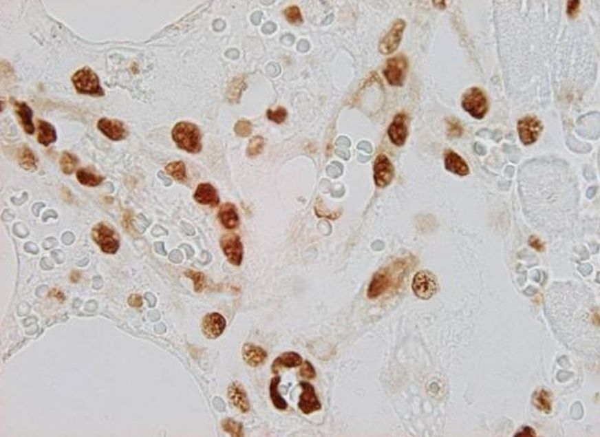
Nestin is expressed in newly formed endothelial cells of blood capillaries, i.e. endothelial cells expressing nestin also co-express proliferative markers (e.g., PCNA). For that reason nestin can be identified in blood vessels of developing organs not only in intraembryonic but also in extraembryonic tissues including placenta (Mokrý and Němeček, 1998 – see Fig. 10). In adult tissues which have fully developed blood vessels nestin can be detected in transplants, e.g. neural ones, which are vascularized by ingrowing blood vessels (Mokrý and Němeček, 1999). Similarly nestin can be identified in vascular endothelium of growing tumours and also in physiologically growing tissues like in the corpus luteum or endometrium. Nestin-positive vessels also appear in regeneration. In patients after myocardial infarction nestin-immunoreactive blood vessels are growing toward the ischaemic tissue (Mokrý, J. et al. 2004 - Fig. 11).
 |
 |
|
| Fig. 10. Immunohistochemical detection of nestin in blood vessels of placenta. Newly formed blood vessels in chorionic villi of human placenta (left image) are lined with nestin-positive endothelium (brown) while erythrocytes and section background are not stained. In rat placenta chorionic villi contain small and large blood vessels (right image); lumen of large blood vessels is indicated by asterisk. Immunoperoxidase reaction; counterstained with light green. Author of microphotograph: Jaroslav Mokrý. |
||
 |
 |
|
| Fig. 11. Immunohistochemical detection of nestin in v regenerating blood vessels after myocardial infarction. Immunofluorescent detection of nestin (left image - red) in the cytoplasm of endothelium of newly formed blood vessels. Nuclei of nestin-positive endothelial cells expressing PCNA (right image - brown) provide evidence of newly formed cells. Author of microphotograph: Jaroslav Mokrý. |
||
