
PSEUDOSTRATIFIED EPITHELIA
Interpretation of microscopic structure and examples of pseudostratified epithelia.
2. PSEUDOSTRATIFIED COLUMNAR EPITHELIUM
|
Pseudostratified columnar epithelium consists of both lower basal cells (often triangular in section) and tall columnar cells (i.e. cells of different heights). The cell nuclei are therefore distributed in multiple rows. However, all cells are anchored to the basal lamina. The most common example (Fig. 2) consists of one row of basal cells and one row of columnar cells. The columnar cells of multi-row epithelia may then have a free apical surface (Fig. 2, right) or may bear stereocilia (Fig. 2, left) or kinocilia (Fig. 4). Fig. 2. Schematic drawing of a pdesustratified columnar epithelium with and without stereocilia. Legend is given in the text; the basal lamina is shown in magenta. |
|
|
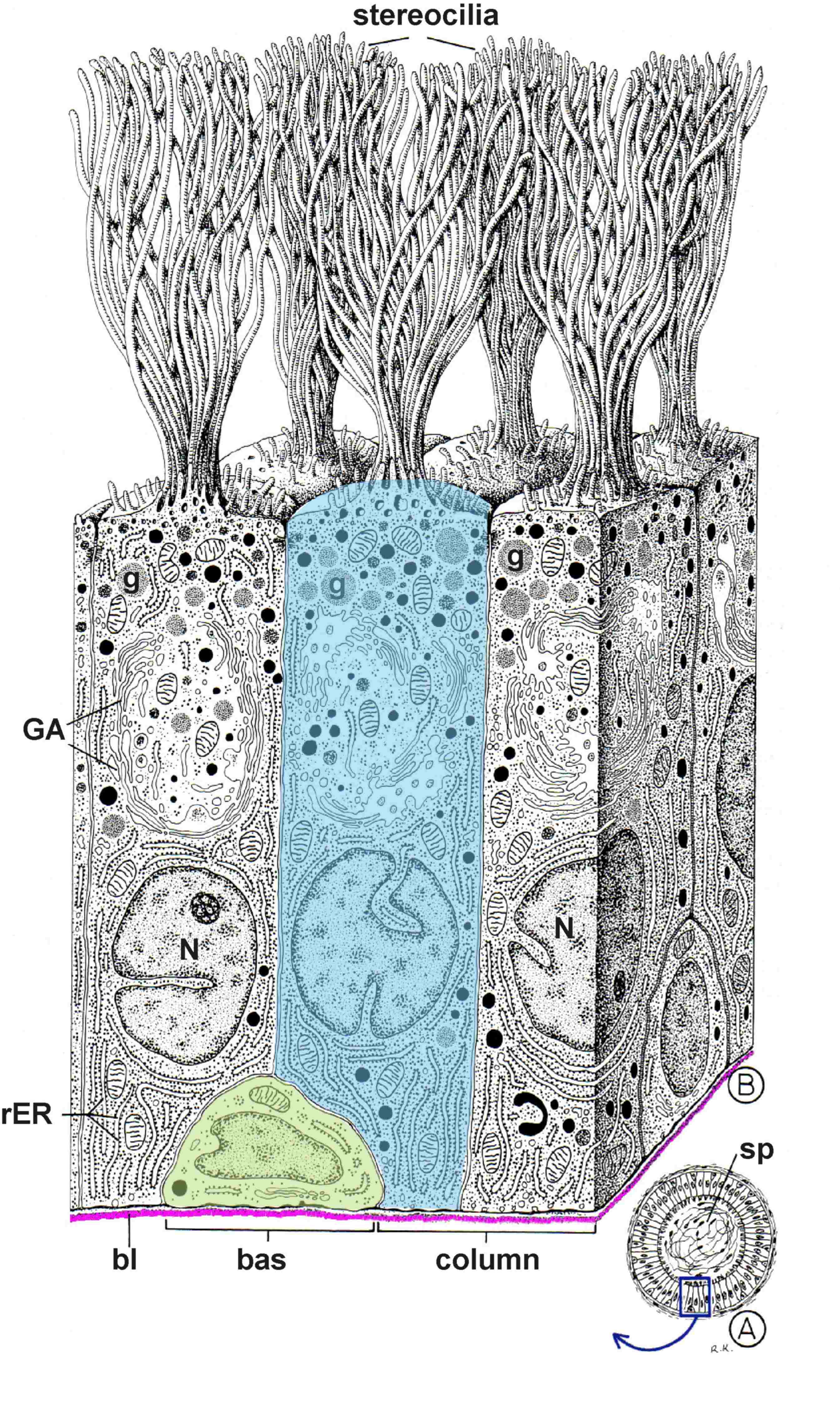
Ductus epididymidis The ductus epididymidis is lined by two rows of epithelial cells (B), which line the lumen of the duct (A), which is filled by spermatozoa (sp). All cells of the covering epithelium sit on the basal lamina (shown in magenta), although they do not all reach the surface of the lining and their nuclei (N) are therefore arranged in different rows. The basal cells (green) are low, while the second row consists of tall columnar cells (blue) with stereocilia. Basal cells have only poorly represented organelles - the Golgi apparatus (GA), secretory granules (g), granular endoplasmic reticulum (rER), mitochondria, lysosomes and lipofuscin granules can be found in the cytoplasm of columnar cells.
Fig. 3. Three-dimensional reconstruction of the epithelial lining of the ductus epididymidis. Legend is given in the text to the left of the figure. |
 |
|
A pseudostratified columnar epithelium with cilia lines the respiratory tract and is therefore referred to as the respiratory epithelium. The respiratory epithelium is taller and contains more rows of cells and its cells vary significantly in height - but all cells are anchored to the basal lamina (magenta). Under the cilia, a dark line is clearly visible under light microscopy by the accumulation of the basal bodies. Another characteristic feature of respiratory epithelia is the presence of goblet cells (unicellular intraepithelial glands) producing mucus (normal staining does not stain the mucus and therefore these cells appear bright). Fig. 4. Schematic drawing of a pseudostratified columnar epithelium with kinocilia and goblet cells. Legend is given in the text; the basal lamina is shown in magenta. |
|
|
Trachea
All epithelial cells sit with their (even if very thinned) base on the basal lamina (in magenta), but only some of them (fully differentiated cells) reach the free (luminal) surface of the epithelium. Thus, the cell nuclei are arranged in multiple rows, which is a basic characteristic of a pseudostratified epithelium. The low basal cells of the epithelium (green) are poor in organelles. They are a cellular reservoir and give rise to about 2% of the tall columnar cells with kinocilia that die each day. The second type are spindle-shaped, narrow elements (purple) with apices in the middle zone; their cytoplasm is richer. They represent an intermediate stage in the differentiation of tall columnar cells from basal cells. This type of epithelium is characterized by tall ciliated columnar cells (shown in red) with kinocilia (cilia) and corresponding basal bodies - kinetosomes (kin). The cytoplasm of the cells is rich in organelles. Because of their high specialization, these elements have lost the ability to divide and must differentiate, as shown above, from the basal cells. In addition, glandular goblet cells (stained blue) secreting mucus are present in the epithelium. The amount of secretory granules (g) in the goblet cells varies according to the functional state. The granules released from the goblet cells mix with the secretion of the serous glands of the respiratory tract. A thin layer of mucus is formed (not shown) which moistens the luminal surface of the epithelium and traps most of the dust particles that enter the airways with the inhaled air. The oscillating movement of the cilia of the respiratory epithelium transports the dust and mucus into the larynx and then into the pharynx, where it is removed during coughing. The epithelium also contains scattered endocrine cells (cells of the diffuse neuroendocrine system, DNES - yellow), which, like the surrounding epithelial cells, touch the basal lamina (magenta). Fig. 5. Three-dimensional reconstruction of the respiratory epithelium of the trachea. Legend is given in the text to the left of the figure. |
 |

