DORSAL ROOT GGL.
2. SLIDE
|
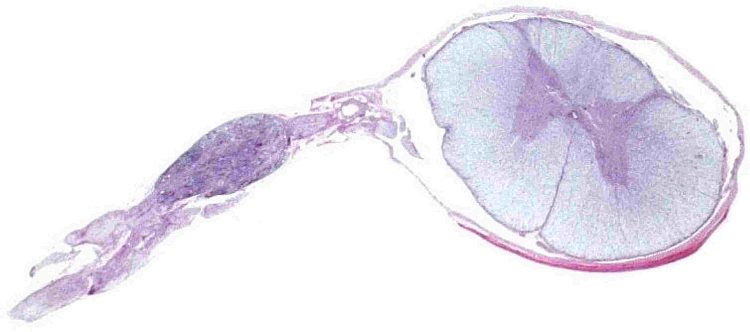
ORIENTATION IN THE SLIDE Slide of the spinal cord of a pig (in an interactive slide indicated in yellow) shows a transverse section that also includes the dorsal root ganglion (in green). In the spinal cord a buterfly-like structure of the grey matter can be seen; well visible are also ventral horns that are shorter than dorsal horns reaching a dorsal surface of the spinal cord. From outside the grey matter is surrounded by the white matter. In the centre the central canal is well apparent. Below is the median anterior fissure filled with a loose connective tissue derived from the pia mater. Coverings of the spinal cord (brown) are continuous with coverings of the roots, ganglion and nerves. Dorsal root ganglion is inserted in the course of the dorsal roots (dark blue). Fusion of the ventral root (not indicated in any colour but it is visible under the ganglion) with nerve fibres of the ganglion forms the spinal nerve (indicated in light blue). |
Fig. 2. Interactive slide of the spinal cord with dorsal root ganglion: A survey. Description can be found in the text. Stained with haematoxylin and eosin. |
|
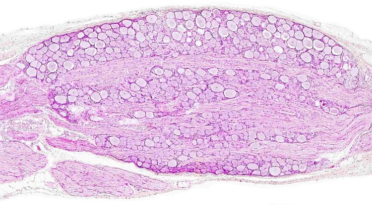
At middle magnification (Fig. 3) large perikarya of ganglionic cells accumulated under the capsule can be found. The capsule (in an interactive slide indicated in brown) is made of the dense connective tissue. Nerve fibres form larger bundles (light green) passing between groups of ganglion cells. These fibres pass through the ganglion to form the spinal nerve (light blue). Histological structure of the ganglion shows that its structural components are well arranged – this is a characteristic feature of all cerebrospinal ganglia. Fig. 3. Interactive slide of the dorsal root ganglion: Middle magnification. Moving a cursor to an image shows structures in the slide in different colours. Description can be found in the text. Stained with haematoxylin and eosin. |
|
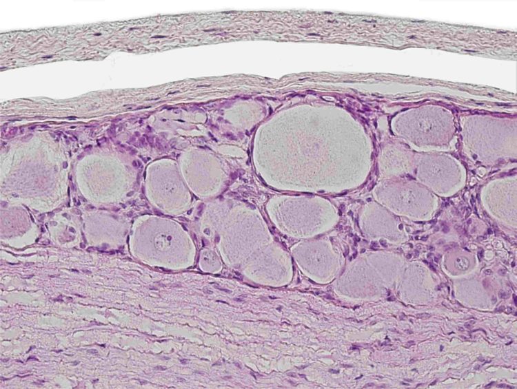
| A detailed microphotograph of the dorsal root ganglion shows a bilayered capsule (brown) made from continuation of the pachymeninx and leptomeninx. Under the capsule a group of ganglion cells (some are labelled with a dotted grey line) can be seen. The neuroplasm contains the well apparent Nissl tigroid substance. Some ganglionic cells contain a spherical nucleus (grey) with a prominent nucleolus (black). A surface of the perikaryon is ensheathed by a layer of satellite cells (nuclei of some apmphicytes is indicated in red). A space visible between the perikaryon and satellite cells is artefactual caused by shrinkage during a tissue processing (dehydration) for histology. Under a group of ganglion cells (black dotted line) nerve fibres can be seen. Size of nerve fibres is different. In a longitudinal section, the axon (orange) can be found inside covered by the white myelin. Ranvier nodes (yellow arrows) are visible as tiny crosses (they are oriented perpendicularly to the axon. Flattened nuclei at the edge of myelin belong to Schwann cells (nuclei of some Schwann cells are indicated in green).
|
 |