ENDOTHELIAL PROGENITOR CELLS
5. INTERMEDIATE FILAMENT NESTIN
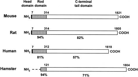
Nestin is a 220 kDa protein (Fig. 7) found in the cytoskeleton of certain cell types; it belongs to intermediate filaments class VI. This protein appears transiently in cell development as it modifies properties of the cytoskeleton and makes it more flexibille. High levels are usually observed in dividing cells. In the course of cell differentiation nestin is changed for another and definitive type of intermediate filament: in neurons nestin is replaced with α-internexin, peripherin or neurofilaments; in astroglia, nestin is replaced with vimentin and GFAP; in muscle cells nestin is replaced with vimentin and later with desmin.
 |
 |
|
 |
||
| Fig. 7. Schematic structure of nestin. Central part of the molecule is conserved (amino acids 7 to 314); helical domain participates in coiling of the intermediate filament. C-terminal part comprises charged amino acids, glutamate residues and motif of 11 repetitive amino acids. Lower image demonstrates interspecies differences in nestin structure. | Fig. 8. Immunoperoxidase detection of nestin in blood vessels of the human corpus luteum. Hormonal stimulation triggers a rapid growth of this endocrine tissue. Its nutrition occurs by newly formed blood vessels, which are lined with ednothelial cells strongly expressing nestin; the section is coverstained with light green. Author of microphotography: Jaroslav Mokrý. |
|
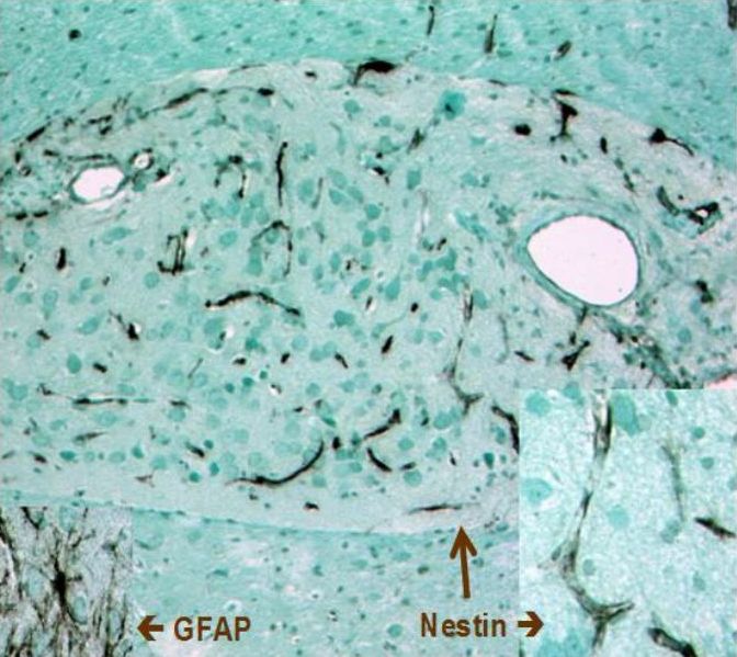
Nestin was originally identified in neuroepithelial stem cells (Lendahl et al., 1990); its name was derived from acronymum NEuroepithelial STem Cell ProteIN. It is very often considered as a marker of neural stem cells, which is not completely right as it is expressed also in other cell types. Nestin is expressed in other neural cells including radial glia, neural precursor cells, oligodendrocyte precursore, premitotic neuroblasts, reactive astrocytes and Schwann cells; of brain tumours nestin was found in gliomas. Nestin appears in developing muscles, in cells of presomitic mesoderm, myotome and also in developing cardiac muscle. Other nestin-positive cells include mesonefric mesenchyma, podocytes, Sertoli cells, odontoblasts or Ito cells. Our group was the first to identify nestin expression in endothelial cells (Mokrý and Němeček, 1998; 1999 - Figs. 8 and 9). Fig. 9. Nestin expression in blood capillaries of neural graft. Angiogenesis of blood capillaries provided nutrition to the transplanted tissue. In 3 weeks after transplantation neural cells differentiated (and therefore they were not containing nestin) whereas endothelium of newly formed blood capillaries express high levels of nestin. Immunoperoxidase reaction of nestin; counterstained with light green.Author of microphotograph: Jaroslav Mokrý. |
 |