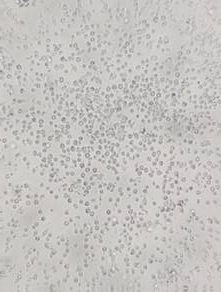
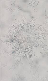
| Mononuclear cells from the blood are centrifuged on Ficoll™ and seeded for 24 hrs in wells coated with fibronectin and containing culture medium EndoCult® Liquid Medium; population of adhering cell is removed. Non-adhering cells are yielded after 2 days and seeded in culture dishes coated with fibronectin. After 3 following days, the first colonies appear (see Fig. 4). Founder cells of the colonies, endothelial progenitor cells, are described as CFU-EC (colony-forming unit-endothelial cells). From 1 ml of blood up to 35,000 endothelial progenitor cells can be isolated. |
 |
 |
 |
| Fig. 4. Isolation of endothelial progenitor cells. Shortly after cell seeding (Fig. In left – 2 days) cell culture contains single cells only. Four days after seeding (middle image) CFU-EC proliferate and give rise to the first colonies that increase their size in the following dates (Fig. In right – 5 days). Photographs were taken from the brochure on Endocult Liquid Medium. |
|
|
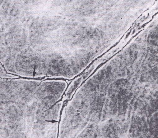
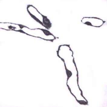
| Endothelial progenitors express specific markers and they also are able to form tubular structures (similar to blood capillaries) in 3D gels (formed e.g. by collagen type I) (Fig. 5). |
|
|
 |
 |
 |
| Fig. 5. In vitro differentiation of endothelial cells. Phase contrast of tubular structures formed by endothelial cells grown in 3D gels (Fig. on left). A central part has a clear lumen whereas edge parts (arrows – middle image) have not been luminized so far. In a semithin section, the endothelium looks like a simple squamous epithelium lining an empty lumen (Fig. on right). |
|
|


